Microscopy

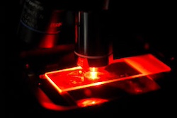
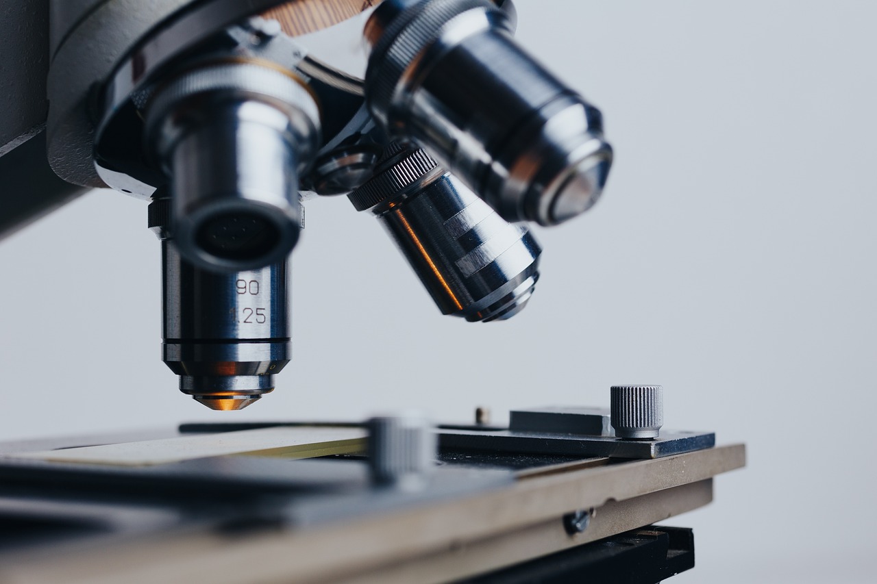
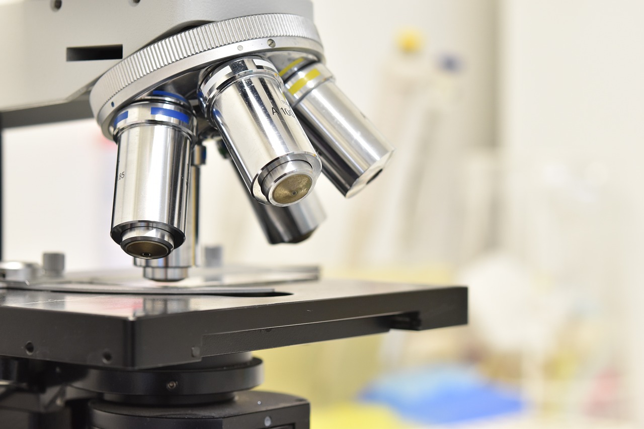
Atomic Force Microscopy (AFM) is a powerful tool to study human pathology, that can work in physiological conditions using different molecules, and cell/ tissue. In this training, participants will be introduced to the AFM  coupled with an Inverted fluorescence microscope, having the opportunity to learn about the determination of morphometric parameters and the characterization of biomechanical properties of cells.

Participants will learn how to use different software for the analysis of the data arising from these studies. Applets software will be used to analyze the force-distance curves, to calculate the mechanical properties of the samples.

Atomic Force Microscopy (AFM) has revealed as a powerful tool to study human pathology, in fields ranging from cancer, cardiovascular and infection diseases, since it is suitable to perform studies on different molecules, cell/ tissue types at physiological conditions. The main principle is based in the interatomic forces established between a probe tip and the sample causing the cantilever to deflect as the samples’ surface topography changes. Laser light reflected from the back of the cantilever measures the deflection of the cantilever and the force applied.

In this lab session, the participants will be introduced to the AFM/IFM techniques, having the opportunity to learn about the determination of morphometric parameters to the characterization of biomechanical properties of cells.

As well, students will learn how to use different software for the analysis of the data arising from these studies. Applets software will be used to analyze the Thermal tune data to obtain the force constant of the cantilevers and to analyze AFM force-distance curves, to calculate the mechanical properties of the samples.